Could a small eyebrow lump be a dermoid cyst?
Finding a lump near your child’s eye is unsettling, so the first thing worth saying is that dermoid cysts are benign. They are not cancer, and they are one of the more common lumps found on a child’s face.
A small lump near the outer eyebrow can certainly fit the pattern, particularly where it has been there since birth or appeared soon afterwards and has grown slowly since. What it actually is, and what if anything should be done, comes from an examination rather than a description.
What it is. A dermoid cyst is a small sac of tissue that became trapped under the skin while your baby was developing in the womb. It can contain hair follicles and glands that produce oil or sweat, and because those glands keep working, the sac can slowly fill and enlarge over years.
Why examination matters. The pattern is recognisable, but a photograph or a description cannot identify an individual child’s lump. What an examination establishes is exactly where it sits, how it relates to the eyebrow and eyelid, and whether anything about it suggests looking deeper.
Where exactly it sits changes everything
“Near the eyebrow” is not precise enough to plan anything. A lump at the outer end of the eyebrow behaves differently from one between the eyebrows or over the bridge of the nose, and the distinction matters more than most parents are told.
| Outer eyebrow pattern | Midline pattern |
|---|---|
| Sits beside the outer end of the eyebrow, sometimes close to the eye socket | Sits between the eyebrows, on the nose, or along the centre of the face |
| Usually lies in superficial tissue, or on the tissue covering the bone | More strongly associated with deeper tracts and connections inside the skull |
| Assessment concentrates on the local anatomy around the eyebrow and eye socket | Assessment has to consider deeper extension carefully before any surgery |
Midline lesions, meaning those between the eyebrows, on the nose or along the centre of the face, are the ones most strongly linked with connections inside the skull. Cysts at the outer eyebrow and around the eye socket usually sit more superficially, though an examination still checks for anything suggesting a deeper connection.
None of that diagnoses a lump from its position. It explains why a surgeon will want to know precisely where it is before discussing anything else.
If you have noticed a mole or lump on your child, it is worth having it looked at. Arrange a consultation
Scans answer questions, they are not a formality
A scan is worth having when it answers something the examination could not. A superficial-looking lump at the outer eyebrow, with reassuring examination findings, may need no imaging before removal, and that is a considered decision rather than a corner being cut.
The assessment runs in order:
- The clinician examines the lump, the skin over it, and how it sits in relation to the eyebrow and eyelid.
- They establish its exact position and how long it has been there, including whether it has been enlarging.
- Those findings decide whether imaging would show something about the deeper anatomy that would change the operation.
Miss Torres-Grau examines each child herself and explains whether imaging is needed before any discussion of treatment.
Magnetic resonance imaging, an MRI scan, shows soft tissue and possible deeper connections, using no ionising radiation. Computed tomography, a CT scan, uses X-rays and shows bone and deeper anatomy well. Which one suits, if either, depends on what the examination found and where the lump lies.
Research on lateral brow cysts supports imaging selectively rather than routinely where a lump looks superficial and nothing on examination suggests otherwise.
A few words from Miss Jana Torres-Grau
Note when the lump first appeared and whether it has changed in size or position since. A cyst present from early on and growing slowly tells a very different story from one that appeared recently.
If surgery is agreed, the scar is planned
A dermoid cyst will not disappear on its own, because it is a sealed sac that keeps filling with a minimal risk of infection. That is the usual reason removal comes up: not because it is dangerous, but because it tends to grow, and a larger cyst in a more awkward position is a harder operation with a worse scar than a smaller one.
The anaesthetic. A general anaesthetic keeps your child asleep and pain-free throughout. The surgeon makes a small incision, removes the cyst as completely as possible, and closes with dissolvable stitches.
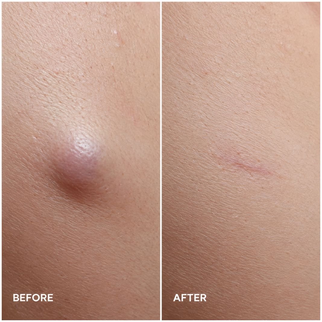
The scar. Where the incision goes is decided at the planning stage rather than in theatre. Depending on the cyst’s position it may sit near the eyebrow or within the eyelid crease, both of which hide a scar better than the alternatives. A scar can still remain visible, and anyone promising otherwise is overselling.
The laboratory. Removed tissue goes for histopathology, meaning examination under a microscope, which confirms what the lump actually was.
Recovery. Swelling and soreness are normal afterwards. Evelina London Children’s Hospital describes a pathway where children usually go home the same day, once they are comfortable, eating and drinking and behaving like themselves. Arrangements vary with the child and the setting.
The recognised risks are bleeding, infection, the cyst rupturing during the operation, recurrence where any lining is left behind, and a visible scar. Follow-up covers how the wound has healed and what the laboratory found.
A few words from Miss Jana Torres-Grau
Be precise about the location: outer eyebrow, between the eyebrows, or over the bridge of the nose. That single detail shapes both the assessment and any decision about scanning.
Common questions about dermoid cysts in children
Can a dermoid cyst go away on its own?
No. It is a sealed sac of tissue rather than a swelling or an infection, so there is nothing for the body to reabsorb. Removal is the only thing that makes it go.
Does every eyebrow dermoid cyst need a scan?
No. It depends on exactly where the lump sits and what the examination shows. A scan earns its place when the surgeon needs a clearer view of deeper tissue, the eye socket or nearby bone, and adds little when they do not.
Will dermoid cyst surgery leave a scar?
There will be a scar, because there is an incision. How noticeable it ends up depends on where it was placed and how your child heals, and placement is planned deliberately around the eyebrow or eyelid crease where the position allows.
Can an eyebrow dermoid cyst return after surgery?
It can, if any of the sac lining is left behind. Complete removal is what reduces that risk, and it is one of the reasons the operation is planned properly rather than treated as a minor skin procedure.
This article gives general information. Only an examination can tell you what is going on for an individual child.
This is general information, not medical advice.
Reviewed by Miss Jana Torres-Grau, Consultant Paediatric Plastic Surgeon, FRCS (Plast), General Medical Council Specialist Register.
A specialist can explain whether anything needs to be done. Ask a question
