
Why has my child been sent for an ultrasound before a lump is removed?
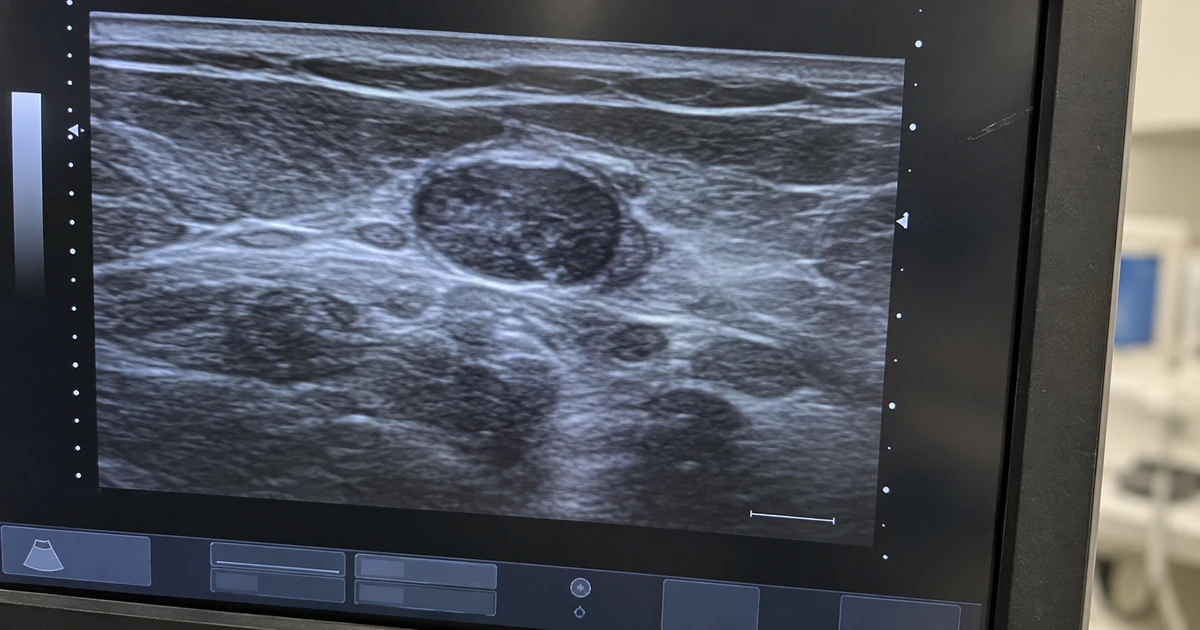
An ultrasound before lump removal gives the surgeon a view beneath the skin. It shows whether the lump appears fluid-filled, solid or mixed, how far it extends, its blood flow and nearby structures. That information may guide the approach to removal or show that more imaging is needed first.
A request for a scan means the surgeon wants more detail before planning.
Examination and ultrasound provide different information
Your child’s examination shows where the lump sits, how it feels and whether it moves under the skin. Ultrasound shows features beneath the surface that hands and eyes cannot assess.
A hand-held probe sends high-frequency sound waves into the tissue. It receives returning echoes, and a computer turns those echoes into an image on a monitor. Ultrasound uses no X-rays or ionising radiation.
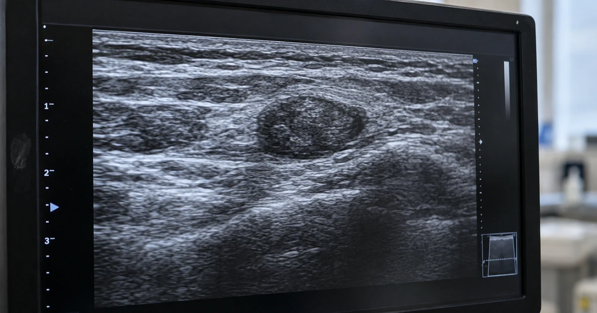
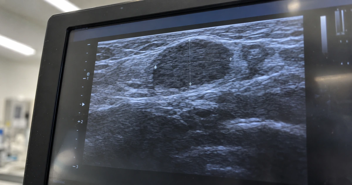
The scan can show whether a lump appears fluid-filled, solid or mixed. It can also show its shape, depth and relationship to underlying structures. Colour Doppler ultrasound is a scan setting that shows blood flow within or around the lump.
The British Medical Ultrasound Society explains that imaging works alongside your child’s history and examination. A 2026 review published in Pediatric Radiology found that different lumps can look alike on a scan, and that key features still narrow the likely diagnosis and help guide the plan.
Lump features guide the decision to scan
A small, mobile lump confined to the skin or tissue just beneath it may give enough information on examination alone. A lump that feels fixed, extends more deeply or lies close to important structures may need ultrasound before removal.
The comparison below shows why two visible lumps can need different preparation.
| Features suggesting examination may be enough | Features that may make ultrasound useful | How the finding could affect planning |
|---|---|---|
| A small, mobile lump that appears confined to the surface tissues | A lump that feels fixed or has uncertain depth | The surgeon can decide whether removal needs a different approach |
| A clearly superficial cyst with a defined edge | A lump near the eyebrow, scalp or facial midline | Imaging can show its relationship to the orbit, skull bone or other nearby structures |
A 2022 review published in the International Journal of Pediatric Otorhinolaryngology found that some lumps around the eyebrow, scalp and facial midline can track towards deeper structures, and that a scan before removal helps where that connection is a concern. Position is one factor, and the surgeon weighs it with how the lump looks and feels.
MRI is a scan that gives detailed pictures of soft tissues. CT is a scan that gives detailed information about bone. Either test may follow when ultrasound leaves a question about depth, full extent or a possible connection through skull bone.
Your child’s consultation covers whether a scan is needed and what question it will answer.
If you have questions about your child’s care, you can arrange a consultation with Miss Jana Torres-Grau. Arrange a consultation
What your child experiences during the scan
A standard child lump ultrasound scan uses gel on the skin and a small probe moved over the area.
During the appointment:
- Positioning: Your child sits or lies in a position that gives access to the lump.
- Gel and probe: The radiology doctor places gel over the skin and moves the external probe across the area.
- Images: The probe sends sound waves into the tissue, and the monitor displays the returning echoes as images.
- Completion: The scan often takes about 10 to 20 minutes. A parent can stay with a young child, and normal activities can resume afterwards.
A standard external diagnostic ultrasound is painless and uses no radiation. No needle is used during a routine scan of a lump.
A separately arranged biopsy or aspiration is different. Those procedures use a needle and would be discussed clearly in advance.
A few words from Miss Jana Torres-Grau
Bring the ultrasound report and images to the consultation if they are available. The report and the images can answer different parts of the surgical planning question.
How the surgeon reads your child’s scan result
An ultrasound result needs to be read with the examination and the history of the lump. The scan provides useful detail, but it cannot answer every question on its own.
A cystic or solid appearance on ultrasound
The lump’s contents (fluid, solid or a mix) help the surgeon narrow down what it is likely to be and plan how to remove it.
When cancer is a worry
Most lumps under the skin in children are benign, as the 2026 review notes. Ultrasound is not used on its own to exclude cancer, because the British Medical Ultrasound Society, in its guidance on children’s neck lumps, notes that harmless, infected and cancerous lymph nodes can share some scan features. The surgeon reads the scan with the examination and arranges further tests where a question remains.
How ultrasound assesses depth
A scan shows how deep a lump goes and how far it extends. A deep lump or one with an unclear edge may need MRI or CT to answer the remaining surgical question.
Possible attachment to the skull
The images show how a lump sits against nearby structures. Where a possible connection through skull bone remains unclear, CT or MRI provides the detail needed before removal is planned.
Further imaging is used when the ultrasound leaves an important question about surgery unanswered.
A few words from Miss Jana Torres-Grau
Ask whether the scan has answered the question about depth and nearby structures. If it has not, the surgeon can explain whether further imaging is needed before removal.
Planning after the ultrasound result
Once the report is available, the findings may support what was expected. They may also show that more imaging is needed before an operation, particularly where the full extent of a lump remains unclear.
Miss Torres-Grau examines each child herself and uses the examination with the imaging findings to decide whether surgery is appropriate. She carries out surgery herself when removal is the right step and follows each child through recovery.
Families in London can arrange that assessment directly, and a scan is only requested where it answers a real question about the operation.
Every child is different, so a personal assessment is the best next step. Ask a question
Common questions about child lump ultrasounds
What does fluid-filled mean on an ultrasound report?
Fluid-filled describes how a lump appears on the scan. It gives the surgeon information about its contents, but the final diagnosis may still depend on examination or tissue analysis after removal.
What does colour Doppler ultrasound show?
Colour Doppler adds colour to the image where blood is moving. It shows the surgeon how much blood supply a lump has, which helps plan its removal.
Who performs a child lump ultrasound?
In Evelina a radiology consultant performs and reports the ultrasound examinations. Sometimes this will be done together with Miss Torres-Grau’s examination.
Does an ultrasound result always mean my child will have surgery?
No. The result may support removal, lead to further imaging or show that surgery is not the right immediate step. An examination and the scan findings define the plan.
Do I need a GP referral for a private consultation?
No. You can arrange a private consultation directly. A GP referral is useful when you already have one because it provides background information about your child.
This is general information, not medical advice.
Reviewed by Miss Jana Torres-Grau, Consultant Paediatric Plastic Surgeon, FRCS (Plast), General Medical Council Specialist Register.